The technical problem is obtaining three-dimensional structural information inside of objects in a manner which is nondestructive. The most often applied approach is conventional computed tomography where either the object is rotated around one axis, or the x-ray source and detector are rotated around the object. Then the object structured is determined using a computation algorithm. This limits the application to cases where the object and structure inside the object is not changing over the time period needed to perform the scan. Furthermore, the resolution is determined by the x-ray source and detector characteristics.
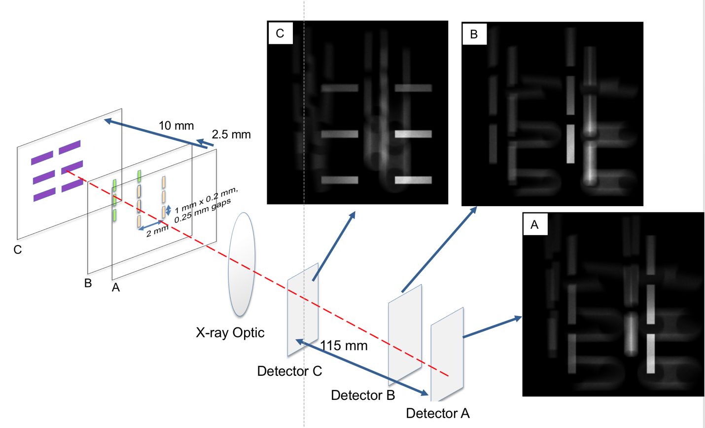
LLNL's 3D X-ray imager combines two different hardware pieces. The first is an x-ray optic with a depth-of-field that is small compared to the object under investigation. Reflective Wolter type x-ray optics are one such design. These hollow optics have a relatively large collection efficiency and can be designed with a large field of view. The depth of focus, which is the distance over which a feature can be resolved along the imaging direction, is relatively small for these optics, typically small compared to the field of view. These optics have been used extensively in x-ray astronomy and in some cases for x-ray microscopy. The short depth of field distance is often considered a drawback to the design. However, when combined with a three-dimensional x-ray detector, it is possible to take advantage of the short depth of field to obtain additional information about the 3D structure of an object. One simple version of the 3D detector uses film. The x-rays are partially transmitted and partially absorbed through a piece of x-ray film. This allows the recording of multiple images along one line of sight and simultaneously. This invention may take advantage of future developments in 3D x-ray detectors that might include thinned CCDs, or CCDs used with x-rays at energies that transmit well through the CCD.
What is novel about this technology is the fact that x-rays are not completely absorbed in a detector and that an optic with a short depth of field can be combined to produce a 3D reconstruction of the object under study.
The analysis of the object will require a back-propagation or similar computational algorithm to take full advantage of the data. The known detector positions allow such a numerical computation of the object under study.
This technology overcomes the limitation of obtaining three-dimensional structural information by providing multiple views with one exposure without moving parts. It relies on a 3D detector, which can be as simple as a stack of film plates, and a focusing x-ray optic. The x-ray optic allows collection of x-rays from a localized volume, just like an ordinary optical lens, and the stacked film plate, or other 3D detector design, allows collection of the multiple focal plane information from one line of sight. Schneberk, Jackson, and Martz suggested using a Wolter optic to acquire multiple images by scanning the Wolter optic and recording multiple images on a single CCD camera (Schneberk, D., J. Jackson, and H. Martz. Possible Laminographic and Tomosynthesis Applications for Wolter Microscope Scan Geometries. No. UCRL-TR-207196. Lawrence Livermore National Lab., Livermore, CA (US), 2004). The limitation is that the object must be stationary during the scan, and the translation of the optic needs to be done so as to not introduce additional artifacts. By acquiring multiple images from a single line of sight with a 3D detector, the time dependence is removed from the imaging.
Furthermore, acquiring images simultaneously from a single line of sight reduces the motion blurring and reconstruction artifacts due to the system motion.
This invention may take advantage of future developments in 3D x-ray detectors that might include thinned CCDs, or CCDs used with x-rays at energies that transmit well through the CCD.
One use of this technology is in imaging extended laser-produced x-ray sources where high resolution and high throughput is required over an extended volume. A second use would be imaging objects small compared to the depth-of-field of the microscope, with the multiple views allowing one of the images to be in-focus. This could be applied to medical imaging to allow determination of the three-dimensional structure without the complication of rotating the x-ray source and detector used in typical computed tomography radiography. A single line-of-sight with the three-dimensional detector and short depth-of-focus optic allows individual slices to be reconstructed to better determine the shape and location of features.
Additionally, other applications that require similar non-destructive evaluation would benefit from such technology. These might include characterization of additively manufactured components, for example, baggage screening, and related applications. Applications where inspection with a computed tomography system is impractical may be enabled by this technology. For example, inspection of components in the field, or small regions inside of a larger part may benefit from this technology.
Additionally, this concept will be applicable to other penetrating radiation that can be focused, such as low-energy neutrons.
A laboratory prototype has demonstrated imaging using Mo K-alpha radiation, often used in mammography, with approximately 0.25 mm lateral resolution and 4 mm depth resolution.
LLNL has filed a patent application for this invention, U.S. Patent Application No. 16/174625 (IL13229).


