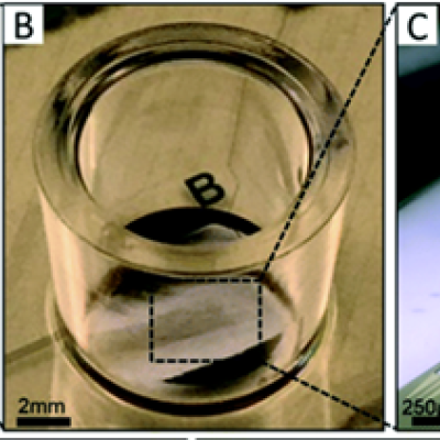
To replicate the physiology and functionality of tissues and organs, LLNL has developed an in vitro device that contains 3D MEAs made from flexible polymeric probes with multiple electrodes along the body of each probe. At the end of each probe body is a specially designed hinge that allows the probe to transition from lying flat to a more upright position when actuated and then…
Keywords
- Show all (121)
- Instrumentation (38)
- Additive Manufacturing (37)
- Diagnostics (13)
- 3D Printing (7)
- Therapeutics (5)
- Brain Computer Interface (BCI) (3)
- Manufacturing Improvements (3)
- Synthesis and Processing (2)
- Vaccines (2)
- Electric Grid (1)
- Manufacturing Simulation (1)
- Material Design (1)
- Microfabrication (1)
- Precision Engineering (1)
- Rare Earth Elements (REEs) (1)
- Sensors (1)
- Volumetric Additive Manufacturing (1)
- (-) Manufacturing Automation (2)
- (-) Polymer Electrodes (1)
Technology Portfolios
Image

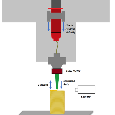
Livermore researchers have developed a method for implementing closed-loop control in extrusion printing processes by means of novel sensing, machine learning, and optimal control algorithms for the optimization of printing parameters and controllability. The system includes a suite of sensors, including cameras, voltage and current meters, scales, etc., that provide in-situ process monitoring…
Image

LLNL researchers have developed a system that relies on machine learning to monitor microfluidic devices. The system includes (at least) a microfluidic device, sensor(s), and a local network computer. The system could also include a camera that takes real-time images of channel(s) within an operating microfluidic device. A subset of these images can be used to train/teach a machine learning…

