
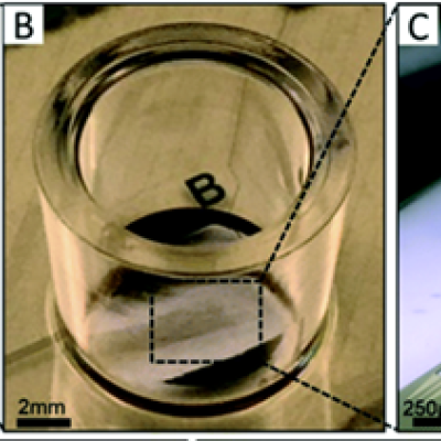
To replicate the physiology and functionality of tissues and organs, LLNL has developed an in vitro device that contains 3D MEAs made from flexible polymeric probes with multiple electrodes along the body of each probe. At the end of each probe body is a specially designed hinge that allows the probe to transition from lying flat to a more upright position when actuated and then…
Image
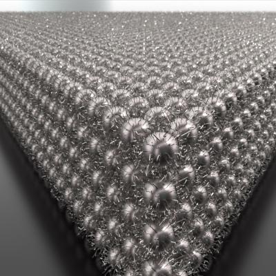
The novel LLNL technique uses electric fields to drive and control assembly. In the literature such methods have heretofore only formed disordered ensembles. This innovative method increases local nanocrystal concentration, initiating nucleation and growth into ordered superlattices. Nanocrystals remain solvated and mobile throughout the process, allowing fast fabrication of ordered…