
Understanding and visualizing the interior landscape of a patient's eye can be critical in both the diagnosis and treatment of ocular diseases. Visual information on the health of key disease and injury indicators such as the optic nerve, blood vessels, and retina can be derived from methods such as optical coherence tomography (OCT). Thus, the more accurate and reliable the OCT results are, the better they can aid optometrists and ophthalmologists in treating a patient. Like most imaging methods, current OCT methods are subject to the twitching and otherwise non-still nature of the human eye, resulting in decreased accuracy and resolution of the tomographic image.
LLNL scientists have developed a method to ensure the accuracy of that tomographic image by applying adaptive optics (AO) to OCT in a single instrument (AO-OCT). AO stabilizes the image being captured by the OCT device by utilizing a Hartmann-Shack wavefront sensor and a deformable mirror, a type of mirror designed to compensate for detected waveform abnormalities (such as ones caused by a twitching eye) to produce a constant waveform image resulting in a sharp instead of a blurry image. The lateral resolution of the image is further improved by means of a second deformable mirror. Together, the sensors and mirrors stabilize and sharpen the image using the same light source as the OCT, where the sensors "instruct" the mirrors to deform to reflect the best image, thus enabling in vivo visualization of 3D cellular features. The AO-OCT system is further improved by insertion of a Badal Optometer and rotating cylinders to correct large spectacle aberrations such as myopia, hyperopic, and astigmatism. LLNL scientists have found this technology to be so effective that even test subjects without any head restraints (e.g. chin rest, head straps) can produce accurate and clear AO-OCT results.
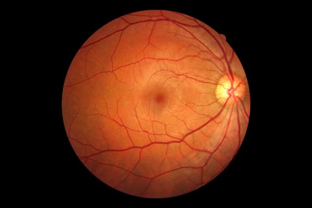

- Produces clear and consistent tomographic images of patients' eyes, specifically of microscopic blood vessels and the cone photoreceptor mosaic
- Use of a single light source reduces the complexity of the invention, improving commercial applicability
- Dual deformable AO mirrors build image quality assurance into the technology
- Real-time processing of AO-OCT images
- Use by optometrists and ophthalmologists in reliably charting the retina and anterior segments of a patient's eye
- Research applications for more clearly understanding how diseases affect the eye
- Use for OCT imaging of other areas of the body
- Veterinary use in non-sedated, non-fully restrained animals
LLNL holds patents 7,942,527 and 8,123,354 both titled "Compact Adaptive-Optical Coherence Tomography System" for this technology (LLNL internal case numbers IL12006A and B), and co-owns a patent 7,791,734 "High-resolution retinal imaging using adaptive optics and Fourier-domain optical coherence tomography" with the Regents of the University of California. (LLNL internal case number IL-11647).