The gold standard in viewing the structure of living cells is light microscopy on a fixed tissue section. However, this requires removal of tissue from the patient ( ex vivo ), and the time required for tissue processing results in significant delays.
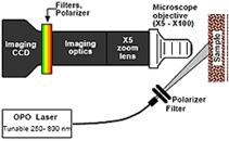
Using various excitation wavelengths, a hyperspectral microscope takes advantage of autofluorescence and polarized light scattering from cellular components to obtain composite images that highlight their presence. The light collection efficiency is maximized to achieve image acquisition times and rates suitable for in vivo applications.
This technology employs the benefits of multi-dimensional spectroscopic imaging at the micro-scale but without the complexity of nonlinear microscopy or the costs, complexity, and signal loss of confocal microscopy.
- Apply in vivo in clinical settings
- Non-invasive to cells
- Provide fast, real-time imaging and monitoring of living cells
- Incorporate a large variety of optical imaging approaches
- Minimal operational complexity
- Can be made portable
- Microscopic imaging tool to assist during surgery
- Monitoring tissues or organs during testing of specific drugs or after exposure to adverse conditions, e.g., radiation
A prototype of the hyperspectral microscope is located at the University of California Davis Medical Center.
The U.S. patent application has been filed for this invention.


