This invention works by imaging an ultrafast pulse diffracted from a large grating onto a spatial light modulator (SLM) thereby directly transcribing an arbitrary record on a pulse front tilted (PFT) ultrafast pulse. The grating generates PFT of the input pulse, and the SLM provides temporal control of the pulse through the space-to-time mapping of the tilted pulse. Coupling this patterned…
Image

This invention exploits the non-linearities of optical Mach-Zehnder (MZ) electrooptic modulators to enhance small signal dynamic range at higher bandwidths. A linear photodiode (PD) converts the amplified optical signal output from the MZ back to an electrical signal completing an Electrical-Optical-Electrical (EOE) conversion cycle. The dynamic range can be further enhanced by daisy chaining…
Image
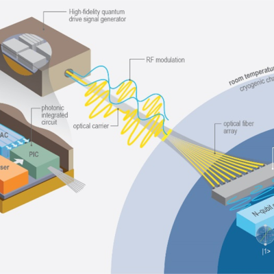
LLNL researchers in the NIF Directorate DoD Technologies RF Photonics Group explored phase modulation solutions to this signal processing challenge. Optical frequency combs offer phase noise characteristics that are orders of magnitude lower than available from commercial microwave references. The Photonics Group researchers recognized that by converting the intensity information into phase,…
Image
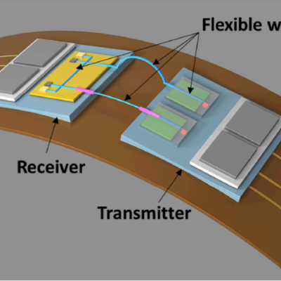
Commercial fiber optic cables are the current standard for carrying optical signals in industries like communications or medical devices. However, the fibers are made of glass, which do not have favorable characteristics for applications that require flexibility and re-routing, e.g. typically brittle, limited selection of materials, dimension constraints.
Image
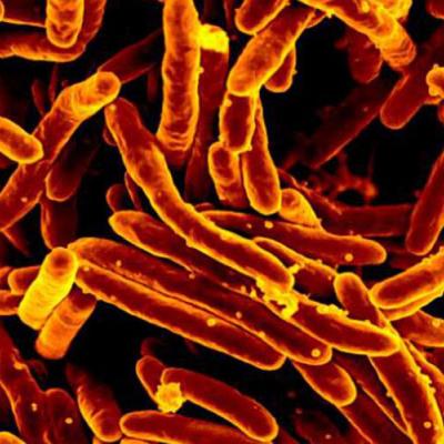
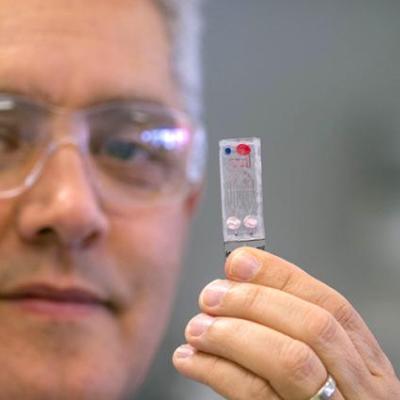
LLNL researchers have developed a high-volume, low-cost diagnostic test that is easy to use and provides results in under an hour. The testing platform will provide emergency responders and other medical professionals with the ability to screen individuals using oral and nasal samples, and obtain results in approximately 30 minutes. This point-of-care testing approach will enable rapid triage…
Image
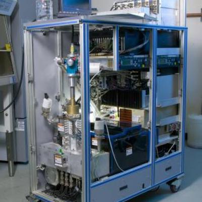
LLNL scientists have created a standalone pathogen identifier that can be placed in public settings, such as in stores or on street corners. Not unlike an ATM in physical size, this kiosk will accept biological samples from an individual for multiplexed analysis. The sample collection process will be sufficiently simple such that anyone could begin the diagnostic process after making the…
Image

LNLL scientists have invented a method for multiplexed detection of PCR amplified products which can be completed in a single step. Highly validated species-specific primer sets are used to simultaneously amplify multiple diagnostic regions unique to each individual pathogen. Resolution of the mix of amplified products is achieved by PCR product hybridization to corresponding probe sequences,…
Image

This LLNL-developed invention is multiplexed and utilizes the Luminex bead-based liquid array, which contains 100 different unique beads. Oligonucleotide probes with sequences complementary to the target sequences are covalently coupled to these unique beads. These capture beads are mixed with viral samples obtained from the patient via cheek swabbing or a throat wash and subjected to PCR in a…
Image

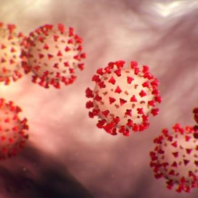
LLNL scientists have developed a high-confidence, real-time multiplexed reverse transcriptase PCR (RT-PCR) rule-out assay for foot and mouth disease virus (FMDV). It utilizes RT-PCR to amplify both DNA and RNA viruses in a single assay to detect FMDV as well as rule out other viruses that cause symptoms in livestock indistinguishable from those caused by FMDV, such as Bovine Herpes Virus-1 (…
Image

LLNL researchers have invented a system for identifying all known and unknown pathogenic or non-pathogenic organisms in a sample. This invention takes a complex sample and generates droplets from it. The droplets consist of sub-nanoliter volume reactors which contain the organism sized particles. A lysis device lyses the organisms and releases the nucleic acids. An amplifier then magnifies the…
Image
LLNL researchers have developed a portable device which analyzes one or multiple types of body fluids or gases to test for one or more medical conditions. A bodily fluid (such as blood, perspiration, saliva, breath, or urine) is put into a condenser surface and is then separated into both a primarily gas fluid component and a second one that is primarily liquid. These two samples from the same…
Image
LLNL researchers have developed a method to quickly and accurately identify the family of a virus infecting a vertebrate via PCR. Universal primer sets consisting of short nucleic acid strands of 7 to 30 base pairs in length were created to amplify target sequences of viral DNA or RNA. These primers can amplify certain identifying sequences of all viral genomes sequenced to date as well as…
Image

LLNL scientists have developed a battery-powered device which is low-cost and multi-chambered for the extraction and amplification of nucleic acids from environmental, clinical, and laboratory samples via loop-mediated isothermal amplification (LAMP). This platform identifies pathogenic bacteria and assists in determining the optimal treatment plan. A multi-chamber amplification cartridge in…
Image
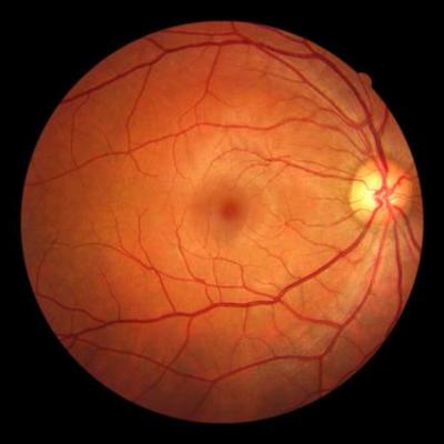
LLNL scientists have developed a method to ensure the accuracy of that tomographic image by applying adaptive optics (AO) to OCT in a single instrument (AO-OCT). AO stabilizes the image being captured by the OCT device by utilizing a Hartmann-Shack wavefront sensor and a deformable mirror, a type of mirror designed to compensate for detected waveform abnormalities (such as ones caused by a…
Image
Using various excitation wavelengths, a hyperspectral microscope takes advantage of autofluorescence and polarized light scattering from cellular components to obtain composite images that highlight their presence. The light collection efficiency is maximized to achieve image acquisition times and rates suitable for in vivo applications.
Image
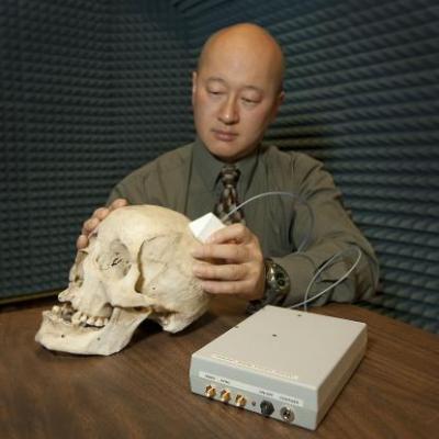
The patented intracranial hematoma detection technology uses Micropower Impulse Radar (MIR). MIR uses short, high frequency electromagnetic pulses to obtain information in a non-invasive manner. Unlike ultrasound and other electromagnetic techniques, MIR can operate well through the skull, which is of great importance for intracerebral as well as epidural and subdural hematomas. The MIR…